Wrist Bone Anatomy
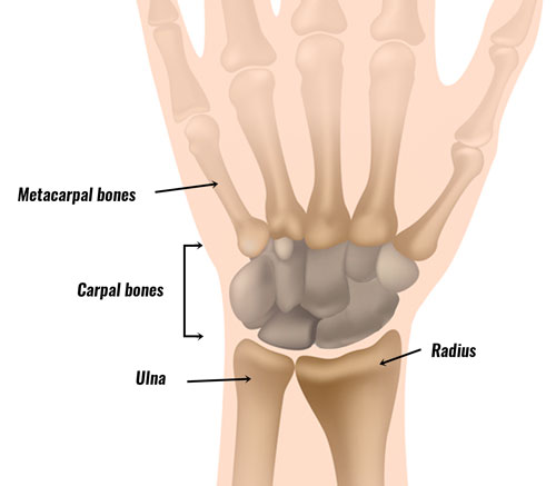
The bones of the hand and wrist provide the body with support and flexibility to manipulate objects in many different ways. The ulna is the arm bone that ends at the wrist on this side.

Carpal Bones Anatomy How To Relief
Tutorial On The Anatomy Of The Wrist Bones And Ligaments News


Wrist Anatomy
This joint is where the radius one of the forearm bones joins with the first row of wrist bones scaphoid lunate and triquetrum.
Wrist bone anatomy. There are two long bones in the forearm that run from the elbow to the wrist. All the joints involving the carpal bones are synovial joints where the articulation surface has a flexible cartilage layer along with a fluid lining to allow for better freedom of movement 22. The wrist joint is a complex joint which connects the forearm to the hand allowing a wide range of movement.
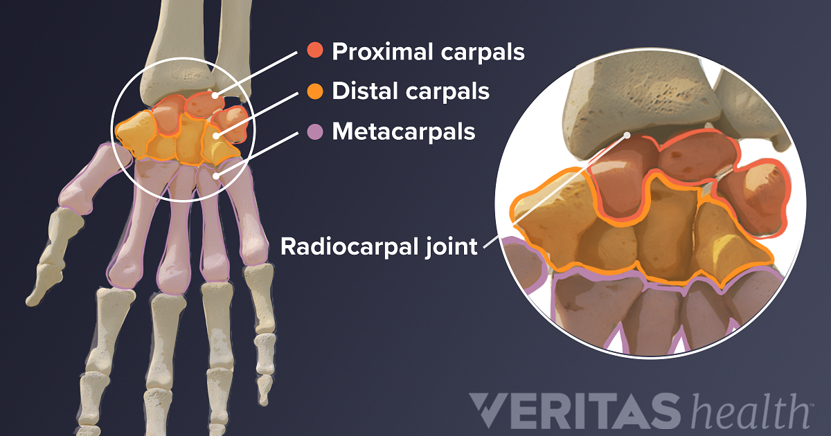
There are three joints in the wrist. Those between the radius and the proximal carpal bones except pisiform 8. The bones in and around the wrist consist of the forearm bones carpal bones and hand bones.
The scaphoid and the lunate are the two bones which actually articulate with the radius and ulna to form the wrist joint. Wrist sprain is an injury to any of the ligaments which connect bone to bone in the wrist. Non-contrast axial CT head.
A fall onto an outstretched hand can lead to a scaphoid fracture. Many wrist injuries such as fractures also known as a broken bone involve the joint surface. Non-contrast coronal CT head.
The hyoid bone is a U-shaped bone that is held in place by the strap muscles of the anterior triangle of the neckThe bone has a central body forming the center of the U with two smaller protruding structures on the superior surface lesser horns and two larger bony protrusions from the body greater horns. The palmaris longus tendon is a tendon with very little function in the hand. The distal radioulnar joint which acts as a pivot for the forearm bones.
Rather than a single joint the wrist is actually made up of multiple joints where the bones of. Non-contrast sagittal CT head. Ulnar wrist pain occurs on the outside pinkie-finger side of your wrist.
The scaphoid bone crosses both rows as it is the largest carpal bone. Therefore the radius is considered to be the larger of the two. It consists of the distal ends of the radius and ulna bones eight carpal bones and the proximal ends of five metacarpal bones.
Wrist anatomy is the study of the bones ligaments and other structures in the wrist. The smaller bone the ulna is on the little finger side. There are different grades of a sprain depending on their severity.
Patients with wrist pain commonly present with an acute injury or spontaneous onset of pain without a definite traumatic event. It attaches to the wrist bone the pisiform and as well as the 5th hand bone. Anatomy of the temporal bone.
The human hand consists of a broad palm metacarpus with 5 digits attached to the forearm by a joint called the wrist carpus. In human anatomy the wrist is variously defined as 1 the carpus or carpal bones the complex of eight bones forming the proximal skeletal segment of the hand. However it is susceptible to injury especially from repetitive strain.
This article lists a series of labeled imaging anatomy cases by system and modality. The tendon travels along the inside of the forearm on the side of the small finger and crosses the wrist. It provides images in the axial and coronal planes allowing the user to review and learn anatomy interactively.
The radius is specially designed to rotate at the elbow and wrist joints around the other forearm bone the ulna. Wrist also called carpus complex joint between the five metacarpal bones of the hand and the radius and ulna bones of the forearm. The wrist is composed of eight or nine small short bones carpal bones roughly arranged in two rowsThe wrist is also made up of several component joints.
Angiogram coronal CT head. What are the signs and symptoms of a medical problem of the ulnar wrist. Ulnar wrist pain while at rest or with movement is a common.
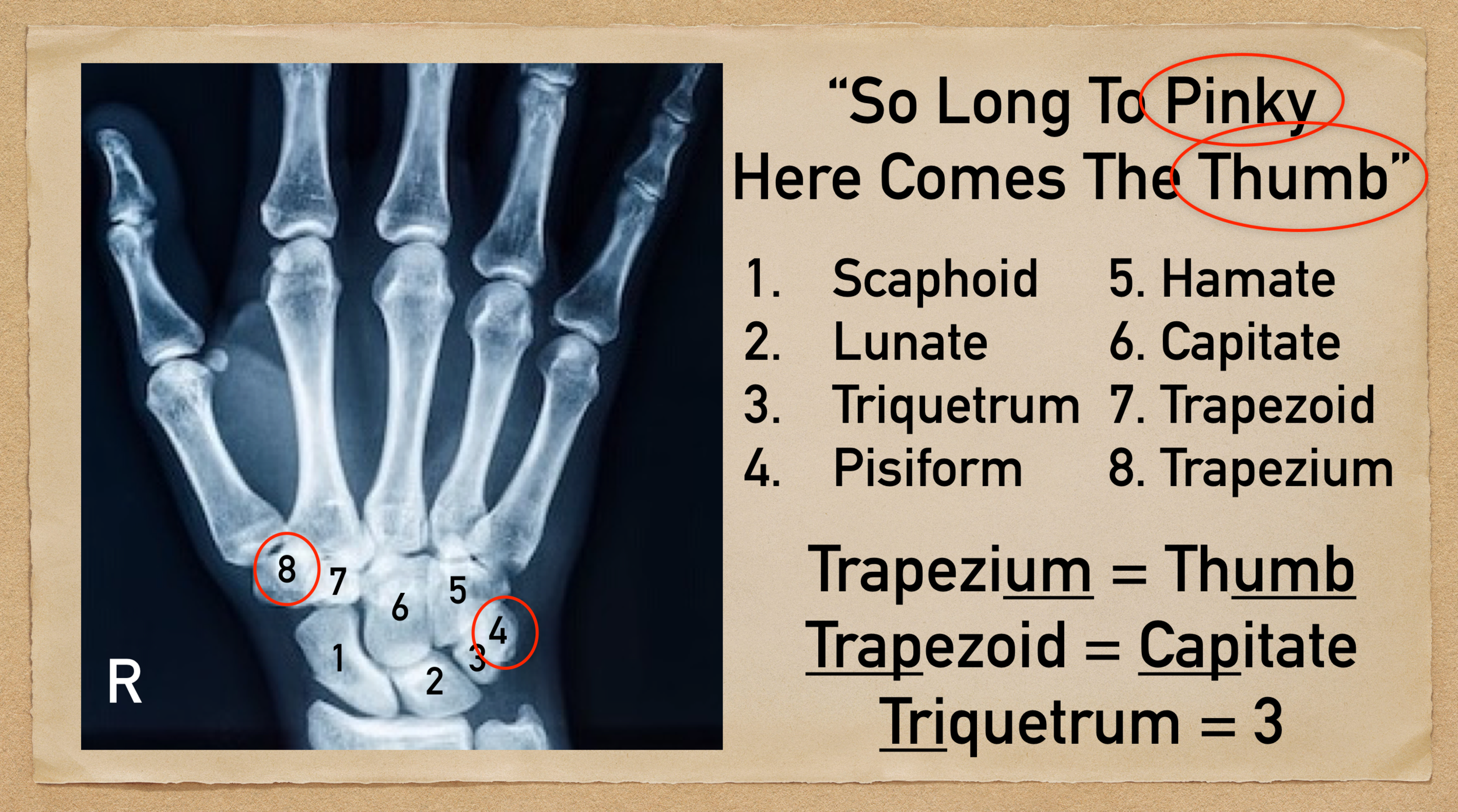
Each wrist is made up of 8 short bones called carpal bones. Scaphoid lunate triquetrum pisiform hamate capitate trapezoid and trapezium. Examples of short bones include the carpal and tarsal bones of the wrist and feet.
In other words the ulna can be found between the proximal carpal row and the upper arm bone humerus running parallel to the other lower arm bone radius 3 5. Knowing the anatomy of the wrist and hand is useful especially when interpreting your own x-rays or performing a physical examination. Radiocarpal joint Articulatio radiocarpalis The radiocarpal joint is a synovial joint formed between the radius its articular disc and three proximal carpal bones.
The ulna is usually slightly longer than the radius but the radius is thicker. It is the medial bone of the forearm located on the side opposite to the thumb that is on the side of the little finger extending from the region of the wrist to the elbow. The carpal bone names are the following.
The wrist is formed where the two bones of the forearm the radius the larger bone on the thumb side of the arm and the ulna the smaller bone on the pinky side meet the carpus. It is a common wrist injury usually caused by a significant impact like a fall. The forearms ulna and radius support the many muscles that manipulate the bones of the hand and wrist.
The radius or radial bone is one of the two large bones of the forearm the other being the ulnaIt extends from the lateral side of the elbow to the thumb side of the wrist and runs parallel to the ulna. This arrangement of. The carpus is formed from eight small bones collectively referred to as the carpal bones.
Allowing the hand to explore and control the environment and objects. Many references however may also include adjacent joints such as the carpal. During pronation the distal end of the radius rotates around the ulna from its position on the lateral side of the wrist to the medial side of the wrist.
A photo taken through a microscope that shows the anatomy of compact bone with a detailed view of an osteon. The wrist is where eight wrist bones two arm bones and five hand bones meet. The end of the long bone is the epiphysis and the shaft is the diaphysis.
Lets take a closer look at wrist anatomy. How to view the anatomical labels. 3 the anatomical region surrounding the carpus including the distal parts of the bones of the forearm and the proximal parts of the.
The scaphoid lunate and triquetral bonesTechnically the radiocarpal joint is considered to be the only articular component of the wrist joint. Angiogram axial CT head. This module is a comprehensive and affordable learning tool for residents and medical students and specially for neuroradiologists and otolaryngologists.
2 the wrist joint or radiocarpal joint the joint between the radius and the carpus and. The larger bone the radius is on the same side as the thumb. The wrist connects the hand to the forearm.
The radiocarpal joint between. Articulations between the carpal bones in hand are an. Bony Anatomy edit edit source The hand and wrist have a total of 27 bones arranged to roll spin and slide 5.
Each hand contains 27 distinct bones that give the hand an incredible range and precision of motion.
Carpal Bone Mnemonic And Names Wrist Anatomy Made Easy Ezmed

Carpal Bones Radiology Reference Article Radiopaedia Org

I Examination Of The Wrist Surface Anatomy Of The Carpal Bones Sciencedirect
Guide To Wrist Anatomy
Left Hand And Wrist Bones Labeled On White Background Stock Photo Download Image Now Istock


Normal Anatomy Of The Carpal Bones Diagram Of The Wrist Frontal View Download Scientific Diagram

Bones In The Wrist Joi Jacksonville Orthopaedic Institute
Wrist Anatomy Bones Ligaments Muscles Nerves
0 Response to "Wrist Bone Anatomy"
Post a Comment