Gram Staining Microbiology Lab
Figure 233 Gram-staining is a differential staining technique that uses a primary stain and a secondary counterstain to distinguish between gram-positive and gram-negative bacteria. Gram stain or Gram staining also called Grams method is a method of staining used to classify bacterial species into two large groups.
Gram Stain Lab Report By Asem Shadid
/microphotograph-of-example-of-staining-bacteria-using-gram-method--at-x1250-magnification-173288072-ab648ac296f846faaa075a7101f06024.jpg)
Gram Stain Procedure In Research And Labs

Pharmaceutical Microbiology Resources Assessing Gram Stain Error Rates Within The Pharmaceutical Microbiology Laboratory
Gram Stain Figure 5.
Gram staining microbiology lab. In the Gram staining protocol two different colored stains can result. Although simple stains are useful they do not reveal details about the bacteria other than morphology and arrangement. Bio 2310L Tablet Visual Guide.
Gram staining is still the cornerstone of bacterial identification and taxonomic division. This is an online free lab manual for the student in BIOL 2310L. Gram-positive bacteria and gram-negative bacteriaThe name comes from the Danish bacteriologist Hans Christian Gram who developed the technique.
The purpose of the Gram stain is to show whether the bacteria are Gram positive purple-colored Gram negative pink-colored or both. 1- introduction Gram Staining 2-Requirements Reagents 3-Method 4-Observations Contents. Dyes are the chemical substances which commonly used to stain specimen.
Microbiology Lab Manual for Face-to-Face Labs. Differential stains use more than one dye. The Gram stain color and the bacterial shape give clues as to what bacteria might be causing the infection.
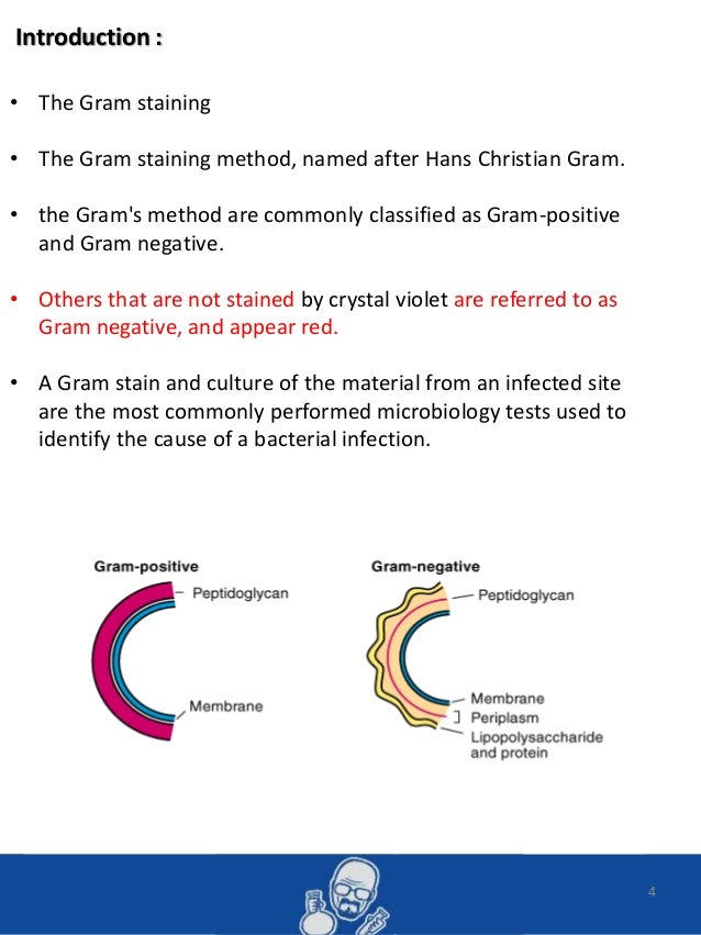
4 The Gram staining The Gram staining method named after Hans Christian Gram. The steps of the Gram stain procedure are listed. It was developed by Danish microbiologist Hans Christian Gram in 1884 as an effective method to distinguish between bacteria with different types of cell walls and even today it remains one of the most frequently used staining techniques.
The Gram stain procedure is a differential staining procedure that involves multiple steps. This method is used for those microorganisms which are not staining by simple or Gram staining method particularly the member of genus Mycobacterium are resistant and can only be visualized by acid-fast staining. There are three types of staining protocol or procedures.
The Gram stain is the most common differential stain used in microbiology. Principle of Acid-Fast Stain. The most widely used staining procedure in microbiology is the Gram stain discovered by the Danish scientist and physician Hans Christian Joachim Gram in 1884.
Gram-positive bacteria retain the color of the primary stain crystal violet in the Gram staining procedure and appear as purpleviolet under a light microscope. Microbiology lab report. Gram positive and Gram negative based on the differences of the chemical and physical properties of the cell wall.
Dark purple staining indicates that the bacteria are Gram-positive and that they have retained the crystal violet stain. It has to be one of the most repeated procedures done in any lab. The Gram stain is a differential stain.
We will be performing the Gram stain and endospore staining procedures in lab and view prepared slides that highlight some of the other cellular structures present in some bacteria. These bacteria have a cell wall containing a thick layer of peptidoglycanOn the basis of cell morphology Gram-positive bacteria are divided mainly into two groups Gram-positive cocci and. The Gram stain is a differential method of staining used to assign bacteria to one of two groups gram-positive and gram-negative based on the properties of their cell wallsIt is also known as Gram staining or Grams method.
Last updated on May 26th 2021. Bacteria stained with Gram stain. Gram staining differentiates bacteria by the chemical and physical properties of their cell walls.
An example of gram-negative bacteria. The purple crystal-violet stained cells are referred to as gram-positive cells while the red safranin-dyed cells are gram-negative. Gram was actually using dyes on human cells and found that bacteria preferentially bind some dyes.
Gram Stain The previous lab introduced simple staining techniques that enable microbiologists to observe the morphological characteristics of bacteria. Gram staining is a differential staining technique that differentiates bacteria into two groups. Gram Stain Summer Term Preparing a Slide with Two Organisms Replaces Two Specimen Slide Video Gram Stain Video.
The gram stain originally developed in 1884 by Christian Gram is probably the most important procedure in all of microbiology. Prokaryotes are identified as gram-positive if they have a multiple layer matrix of peptidoglycan forming the cell wall. The Gram stain is used to classify bacteria on the basis of their forms sizes cellular morphologies and Gram reactions.
In 1884 physician Hans Christian Gram was studying the etiology cause of respiratory diseases such as. The main aim of this staining is to differentiate bacteria into acid fast group and non-acid fast groups. Gram staining bacteria requires the use of aseptic technique to ensure the sterility of the experiment.
The procedure is named for the person who developed the technique Danish bacteriologist Hans Christian Gram. However Gram positive cells may stain Gram negative if they are older or were overexposed to decolorizing agent. One example of gram-positive cocci is Staphylococcus aureus the bacteria associated with staph infections.
For many years the retention of Gram stain was one of. Purple staining indicates a Gram positive reaction and pink indicates Gram negative. If you have mixed purple and pink staining cells that are otherwise indistinguishable then you likely have a Gram positive isolate.
The Gram stain procedure has been basically unchanged since it was first developed in 1884. In contrast reddish-pink staining is a characteristic of Gram-negative bacteria which. The Gram stain is a differential staining technique used to classify categorize bacteria into two major groups.
Materials and Methods Aseptic technique was used throughout the experiment. The Grams method are commonly classified as Gram-positive and. Gram staining differentiates the bacteria into 2 groups.
In a clinical microbiology laboratory it is additionally a critical test for the rapid presumptive diagnosis of infectious. Gram staining bacteria is a fundamental technique introduced in general biology and microbiology laboratory courses. Laboratory perspective of gram staining and its significance in investigations of infectious diseases Yunusa Thairu 1 Idris Abdullahi Nasir 1 Yahaya Usman 2 1 Department of Medical Microbiology University of Abuja Teaching Hospital Gwagwalada Abuja Nigeria 2 Department of Medical Laboratory Science Faculty of Medicine Ahmadu Bello University Zaria Kaduna Nigeria.
HANS CHRISTIAN JOACHIM GRAM The Gram stain was devised by the Danish physician Hans Christian Joachim Gram while working in. Crystal violet the primary stain of the Gram stain procedure is readily retained and stabilized within this matrix causing gram-positive prokaryotes to appear purple under a brightfield microscope after Gram staining. Two common problems students encounter when Gram staining bacteria are 1 having a difficult time locating bacterial cells on the microscope slide and 2 over-decolorizing bacterial cells during the.
Gram staining a differential staining method. The unique cellular components of the bacteria will determine how they will react to the different dyes. Staining is a biochemical technique of coloring specimens.
Gram staining method the most important procedure in Microbiology was developed by Danish physician Hans Christian Gram in 1884. GRAM STAINING Gram staining is most widely used differential staining in Microbiology.
What Is Gram Staining Of Bacteria Stain Is Prepared By Which Material Quora
Gram Negative Bacteria Lab Report Dentalimplantsurgery Com Custom Academic Help

Gram Stain Lab Report Micro Lab

Gram Stain Microbiology Lab
Pdf Development Of A Standardized Gram Stain Procedure For Bacteria And Inflammatory Cells Using An Automated Staining Instrument

Microbiology Lab Exercise 5 Gram Staining Flashcards Microbiology Flashcards Study Flashcards
Gram Stain Lab Questions

Assessing Gram Stain Error Rates Within The Pharmaceutical Microbiology Laboratory
0 Response to "Gram Staining Microbiology Lab"
Post a Comment