Ankle Cartilage Diagram
The ankle is a bit different from the wrist. A high ankle sprain.
Ankle Wikipedia
1
Sprained Ankle Orthoinfo Aaos
Types of Synovial Joints.
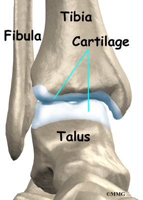
Ankle cartilage diagram. Together they form a bracket shaped socket covered in hyaline cartilage. Ankle sprains and fractures are common sports injuries. Arthritis also can develop from a very severe sprain if the cartilage of.
It is present from birth congenital and is a common traitThe reported incidence differs among populations and ethnic groups and they are mostly reported as incidental findings in anatomical and imaging studies. The inner bone is the tibia or shinbone which supports most of a persons weight when standing. The six types of synovial joints allow the body to move in a variety of ways.
But the main part of the foot is similar to the hand with five bones. A hard outer layer that is dense strong and durable. Because they are the last portion of a childs bones to harden growth plates are particularly vulnerable to injury.
This socket is known as a mortise. This site complies with the HONcode standard for trustworthy health information. A fractured ankle can range from a simple break in one bone which may not stop you from walking to several fractures which forces your ankle out of place and may require that you not put weight on it for a few months.
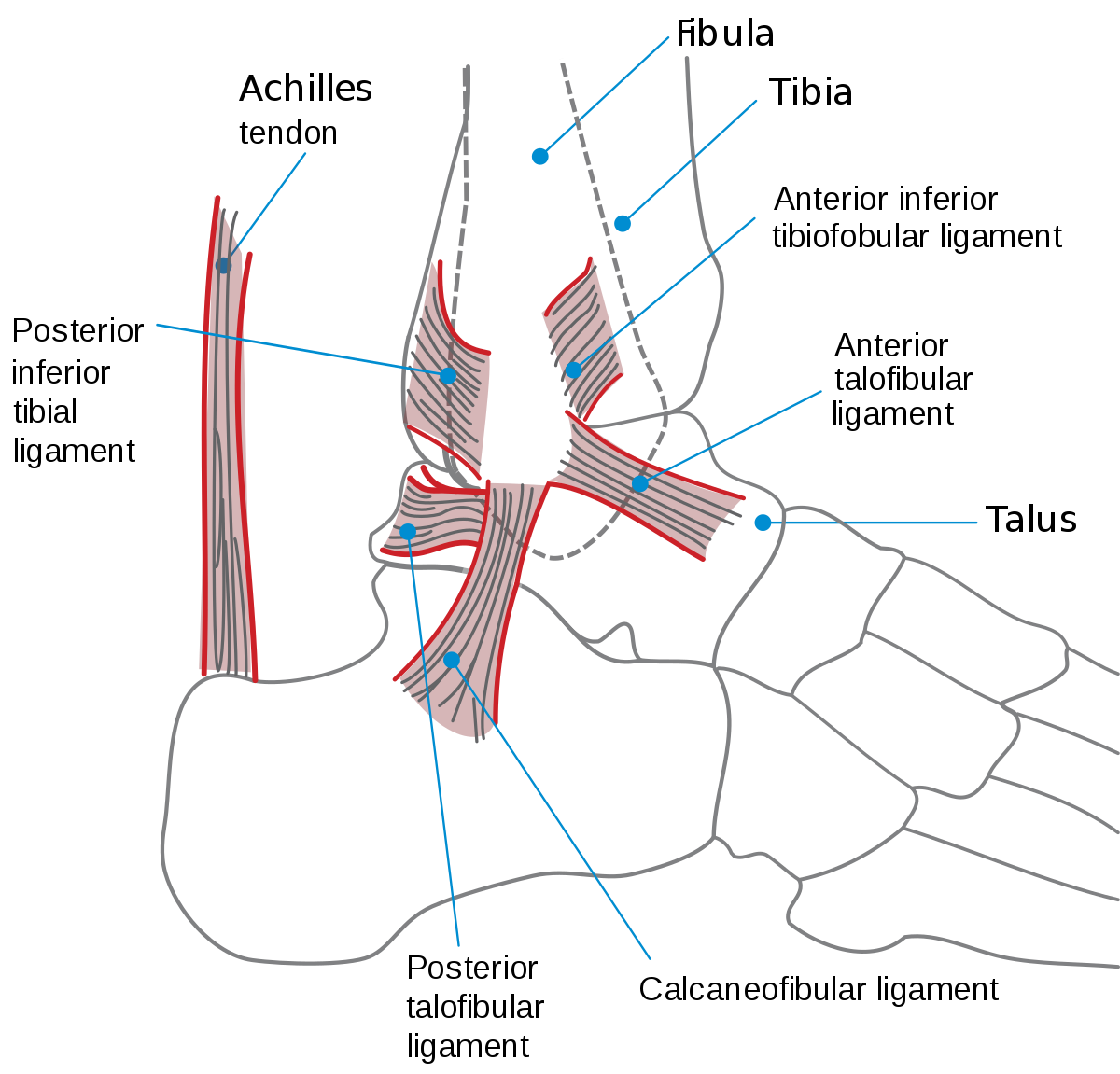
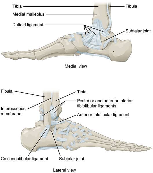
The six types of synovial joints are pivot hinge condyloid saddle plane and ball-and socket-joints Figure 943Figure 943 Types of Synovial Joints. The ankle is a joint that connects the lower leg to the foot. The tibia and fibula are bound together by strong tibiofibular ligaments.
Diabetes mellitus is a very serious metabolic disorder that prevents the normal breakdown and use of food especially sugars carbohydrates by the body. Locate the mammae which are present in both sexes. The outer bone.
Bouchards nodes form on the middle joint of a finger and Heberden. Two types of bony bumps near your finger joints are common. Osteoarthritis is a condition characterized by the breakdown and eventual loss of cartilage in one or more joints.
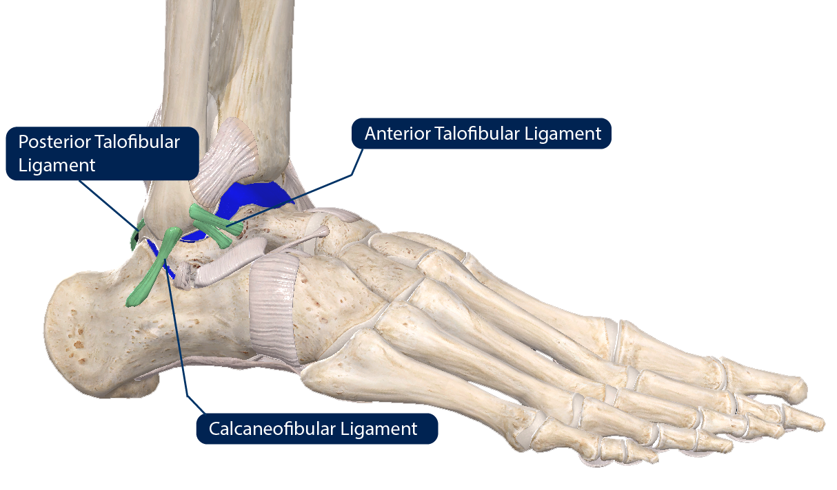
Which join muscles to bone and cartilage which cushions your joints. The ankle joint is formed by three bones. The indications that an ankle sprain has healed are almost as obvious as the initial signs of injury reports Daniel C.
A talar dome lesion is an injury to the cartilage and underlying bone of the talus within the ankle joint. Ankle injuries and ankle disorders can affect tendons and cartilage. List of diabetic foods to eat facts.
A loss of joint space may be due to a loss of cartilage and is commonly seen in conditions such as osteoarthritis. In common usage the term ankle refers exclusively to the ankle region. The ankle is made off the tibia and.
An accessory navicular bone is an extra bone or piece of cartilage located in the middle of the foot near the navicular bone the bone that goes across the foot near the instep. In this event the energy of the injury indicated on the diagram with blue arrows passes from the deltoid through the high ankle. Next to the talus are six other bones.
The Ottawa knee rules are a set of rules used to help physicians determine whether an x-ray of the knee is needed. Each type of cells is specialised to carry out a particular function either solely but usually by forming a particular tissueDifferent tissues then combine and form specific organs where the organ is. They state that an X-ray is required only in patients who have an acute knee injury with one or more of the following.
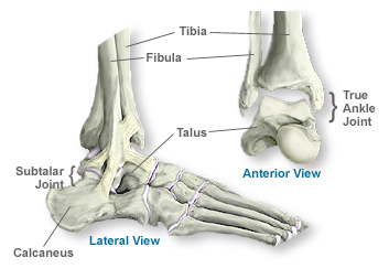
A broken ankle is also known as an ankle fracture This means that one or more of the bones that make up the ankle joint are broken. It is where the lower leg bones connect to a large bone in the foot called the talus say. The tibia and fibula of the leg and the talus of the foot.
Interactive Foot Diagram American College of Foot and Ankle Surgeons X-Ray Exam. But for the patients with sprains that do not heal over time with standard therapy both the cause and next steps for treatment can be unclear. The ankle or the talocrural region is the area where the foot and the leg meet.
Search the Foot and Ankle. Diagram of a mortise on left and tenon joint. The movements produced at this joint are dorsiflexion and plantarflexion of the foot.
Each toe has three tiny. The ankle joint allows dorsiflexion and plantarflexion and is one of the weight-bearing joints. The word sternum originates from the ancient Greek word sternon meaning chest.
It is a flat bone that articulates with the clavicle and the costal cartilages of the upper 7 ribs true ribs while the 8th 9th and 10th ribs false ribs are indirectly attached with sternum via costal cartilage of the ribs above. Osteoarthritis of the Foot and Ankle What Is Osteoarthritis. Calcaneus 2 Talus 2 Navicular bone 2 Medial cuneiform bone 2 Intermediate cuneiform bone 2 Lateral cuneiform bone 2 Cuboid bone 2 Metatarsal Bones.
The ankle includes three joints. Identify the structures on the diagram. The sternum is also known as the breastbone.
Bones are composed of two types of tissue. The ankle joint proper or talocrural joint the subtalar joint and the inferior tibiofibular joint. The tarsal bones are the bones of the ankle and there are 14 tarsal bones 7 on each foot.
Synovial joints are subdivided based on the shapes of the articulating surfaces of the bones that form each joint. They are as under. The ankle is the joint between the foot and leg composed of three separate bones.
It is also called an osteochondral defect OCD or osteochondral lesion of the talus OLT. We would like to show you a description here but the site wont allow us. Growth plates are areas of cartilage located near the ends of bones.
Hand osteoarthritis can cause other problems like. Because the growth plate helps determine the future length and shape of the mature bone this type of fracture usually requires prompt attention. Its main function is to allow for plantar flexion and dorsiflexion of the foot.
Thorax are soft at this stage of development because they are made of cartilage. Cartilage the connective tissue found at the end of the bones in the joints protects and cushions the bones during movement. Most ankle sprains will heal with standard RICE therapy rest ice compression and elevation within two to 12 weeks.
On the inside of the ankle the deltoid will be torn. Locate the wrist and elbow of the forelimb and the knee and ankle of the hind limb. The top of the talus is dome-shaped and is completely covered with cartilagea tough rubbery tissue that enables the ankle to move smoothly.
Once the long bone parts have fused together the only hyaline cartilage left in the bone is found as articular cartilage on the ends of the bone that form joints with other bones. It makes up around 80 percent of adult bone mass. Age 55 years or older.
There are 5 metatarsal bones in each foot one corresponding to each digit. There are over 200 different cell types in the human body. The tibia and fibula form the ankle joint with the talus one of the seven tarsal bones in the foot.
Chronic Ankle Instability Faqs Foot And Ankle Doctor Articles Asia Medical Specialists

Schematic Diagrams Showing Normal Anatomy Of Ankle Cartilage Download Scientific Diagram
Anatomy Of The Ankle Southern California Orthopedic Institute
The Ankle Joint Articulations Movements Teachmeanatomy

Medivisuals The Ankle Medical Illustration

Why Ankle Pain Treatments Chronic Ankle Pain Ankle Joint Pain

Lower Leg And Ankle Foot Joints Lig Cartilage Diagram Quizlet
Ankle Arthritis Eorthopod Com
0 Response to "Ankle Cartilage Diagram"
Post a Comment