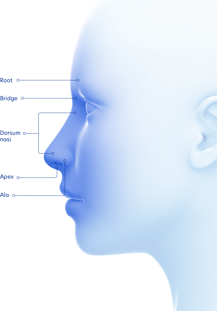
External Nose Anatomy
The external nose is visible and is pyramidal in shape with the root located in the upper region and the base located in the lower region. Their locations and structures are best viewed when the head is shown in sagittal section.

Nose Anatomy Dramatic Context
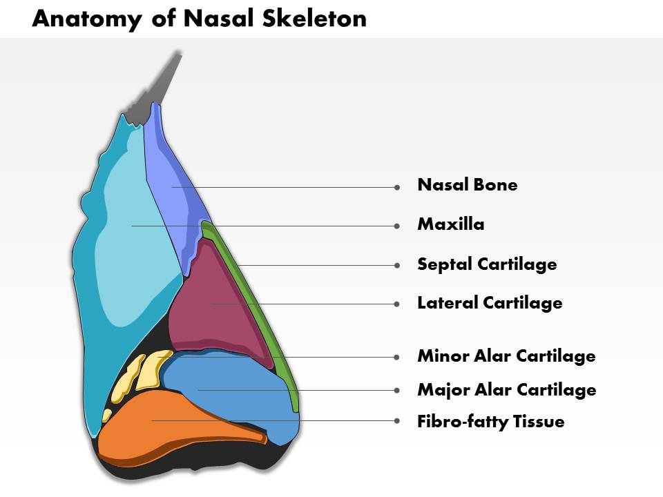
0514 Lateral View Of External Nose Anatomy Of Nasal Skeleton Medical Images For Powerpoint Powerpoint Presentation Pictures Ppt Slide Template Ppt Examples Professional
Anatomy Of The Nose

The site tries to lighten this burden by-providing easy to follow and memorize the concepts of anatomy which are hard to grasp.
External nose anatomy. Anatomy of the Nose. An infection can cause many of the same symptoms as allergic rhinitis. It is subdivided into a left and right canal by a thin medial cartilaginous and bony wall the nasal septum.
Made up mainly of cartilage and bone and covered by mucous membranes. External middle and innerThis article will focus on the anatomy of the external ear its structure neurovascular supply and clinical correlations. The human nose is the most protruding part of the faceIt bears the nostrils and is the first organ of the respiratory systemIt is also the principal organ in the olfactory systemThe shape of the nose is determined by the nasal bones and the nasal cartilages including the nasal septum which separates the nostrils and divides the nasal cavity into two.
Two chambers divided by the septum. The junction of the inferior margin of the nasal ridge and the columella. Anatomists use the Terminologia Anatomica.
The nose is a complex component of the facial anatomy that is comprised of numerous structures. They are also referred to as the vulva or pudendum. The nose helps cats to identify territories other cats and mates.
Triangular-shaped projection in the center of the face. An imaginary line between the most lateral points of the external inferior attachments of the alae nasi to the face. Protruding prominently from the face the nose serves as a vent for air exchange.
Skin fat fascia and muscle. The external nose is a visible component of the face projecting over and allowing entrance into the nasal cavity. Your nose can be broken or injured similar to any other external part of your body.
The advent of endoscopic sinus surgery has increased the interest in the taxonomy of the internal nose and paranasal sinuses. The nose and nasal cavity make up the first portion of the upper respiratory tract. They are divided into the external.
The soft tissue anatomy of the nose can be simplied into 4 layers. The thin superior part that blends with the forehead is called the root of the nose while the region between the apex and the root is called the dorsum. The major landmarks of the external ear are depicted in Figure 1.
Soft Tissue of the External Nose. The external ear consists of skin with adnexa cartilage and six intrinsic muscles. The internal nose is divided into the left and right nasal cavities by the nasal.
In some areas there is. The midline prominence of the nose extending from the nasal root to the tip also called the dorsum of the nose. The auricle or pinna and the external acoustic meatus which ends at the tympanic membrane.
The nose is the external protuberance of an internal space the nasal cavity. Inferior to the apex are the two nares nostrils which are the openings to the nasal cavity. Nose and nasal cavity Sticking out from the middle of your face is your nose a structure that allows you to smell and breatheIt is composed of the nasal bones and cartilage that has two openings called nostrilsBehind the nose is the nasal cavityThere are two cavities in total separated by the nasal septumEach cavity contains three seashell-like structures called nasal conchae and below.
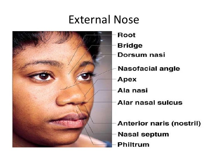
The nasal mucosa also called respiratory mucosa lines the entire nasal cavity from the nostrils the external openings of the respiratory system to the pharynx the uppermost section of the throatThe external skin of the nose connects to the nasal mucosa in the nasal vestibule. Therefore the names used by anatomists and surgeons differ at times. The external part of the nose has a triangular or pyramidal shape with the highest point of it referred to as the apex or the tip of the nose 3.
Here we will go from top to bottom and. Anatomy of the Ear. Examples include sinus infections and the common cold.
The nose is made up of. Learning anatomy can be overwhelming because of the sheer amount of information to be understood and memorized. The external ear can be divided functionally and structurally into two parts.
The first portion of the respiratory tract is made up of the nose or external nose and an open inner chamber called the nasal cavity. Each canal opens to the face by a nostril and into. On each side it is flanked by the maxillary sinuses and roofed by the frontal ethmoid and sphenoid sinuses in an anterior to posterior fashion1 While seemingly simple sinonasal anatomy is composed of.
It is a pyramidal structure with its root located superiorly and apex sitting inferiorlyThe root is continuous with the anterior surface of the head and the part between the root and the apex is called the dorsum of the nose. The bones are called. A dynamic layer of mucus overlies the nasal epithelium the outermost layer of.
Air comes into the body through the nose. The cartilage also gives. They interconnect ribs and are therefore the primary respiratory skeletal muscles.
Anatomy of the nasal mucosa. The nose is the bodys primary organ of smell and also functions as part of the bodys respiratory system. A canal that links the middle ear with the back of the nose.
The tympanic membrane divides the external ear from the middle ear. Its primary function is to protect the lower airway by closing abruptly upon mechanical stimulation thereby halting respiration and preventing the entry of foreign matter into the airway. The external genitalia include the labia majora mons pubis labia minora clitoris and glands within the vestibule.
Lower 13 is less mobile more sebaceous ONeal et al 1999. The anatomy of the domestic cat is similar to that of other members of the. Three small bones that are connected and transmit the sound waves to the inner ear.
Anatomy of the Nose The External Nose. The variance in shape depends on the shape of the ethmoid bone which is an anterior cranial bone located between the eyes. The nasal cavity is a roughly cylindrical midline airway passage that extends from the nasal ala anteriorly to the choana posteriorly1 It is divided in the midline by the nasal septum.
Upper 23 is thin Lessard et al 1985. The external nasal anatomy is quite simple. This article will discuss the anatomy of the external nose its skeletal structure muscles blood supply and innervation.
The larynx is located within the anterior aspect of the neck anterior to the inferior portion of the pharynx and superior to the trachea. The external genitalia are the accessory structures of the female reproductive system that are external to the vagina. Ventilation or breathing is the movement of air through the conducting passages between the atmosphere and the lungsThe air moves through the passages because of pressure gradients that are produced by contraction of the.
The anatomy of the external ear also known as the auricle or pinna is complex Hunter and Yotsuyanagi and remarkably inaccurately described by most authors. The ear can be divided into three parts. Middle ear tympanic cavity consisting of.
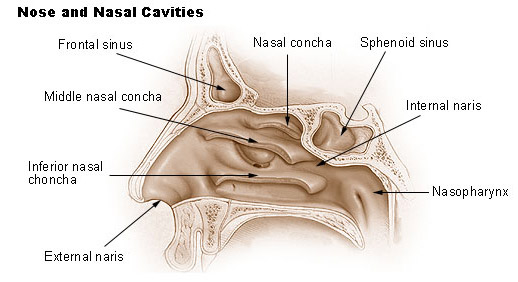
Seer Training Nose Nasal Cavities Paranasal Sinuses
The Nose Anatomy Of The Paranasal Sinuses

0514 Lateral View Of External Nose Anatomy Of Nasal Skeleton Medical Images For Powerpoint Powerpoint Presentation Pictures Ppt Slide Template Ppt Examples Professional
1
Anatomy Of Nose And Paranasal Sinus
Clinical Anatomy Of The Nose
The Nose Contemporary Health Issues


Anatomy External Nose Stock Illustrations 37 Anatomy External Nose Stock Illustrations Vectors Clipart Dreamstime
0 Response to "External Nose Anatomy"
Post a Comment