Hand Bone Anatomy
The scaphoid bone of the hand is the most commonly fractured carpal bone typically by falling on an outstretched hand FOOSH. This thickness also allows the bone to remain strong and sturdy to protect the more delicate features of the face.

Hand Bones Anatomy In Detail

Hand And Wrist Bones Human Anatomy Vector Sketch Hand And Wrist Bones Vector Sketch Of Human Anatomy And Medicine Design Canstock

Hand Anatomy Overview Summit Orthopedics Guide
The upper limb has sacrificed locomotor function and stability for mobility dexterity and precision.
Hand bone anatomy. The anatomy of the hand is incomplete without understanding the wrist. Intrinsic plus hand is a contracture of the intrinsic hand muscles characterized by excessive flexion at the metacarpophalangeal MCP joints and extension at the interphalangeal IP joints. The structures that form and move the hand require proper alignment and control in order for normal hand function to occur.
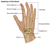
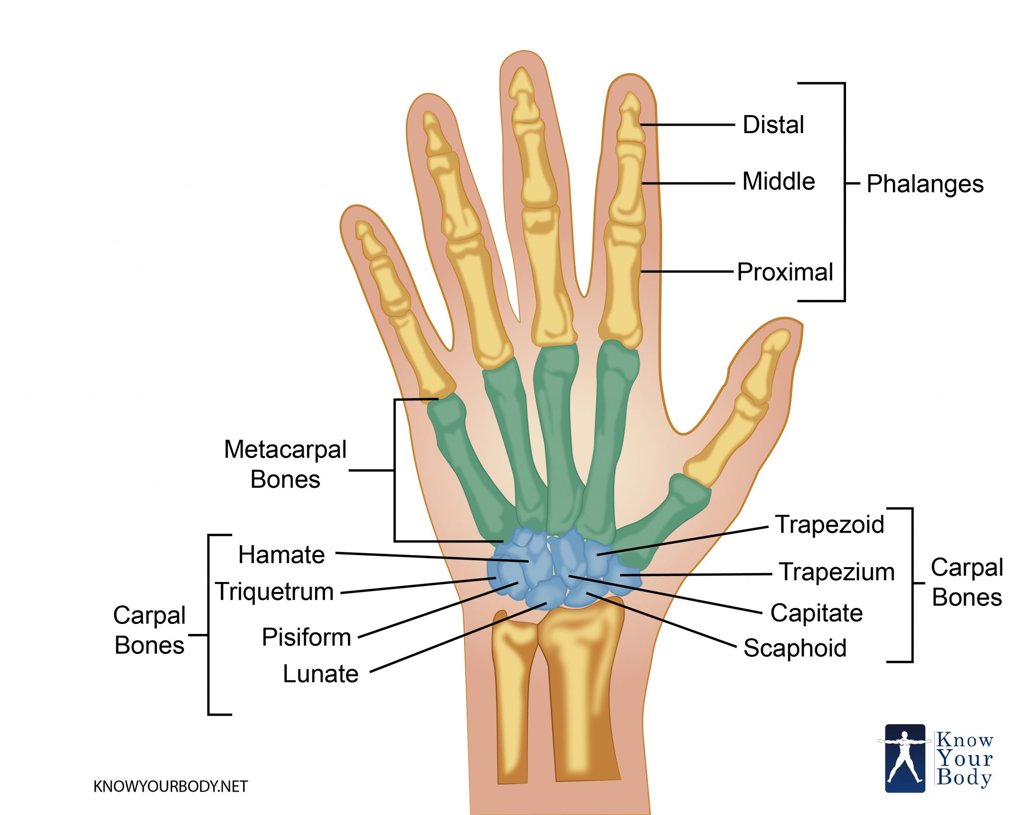
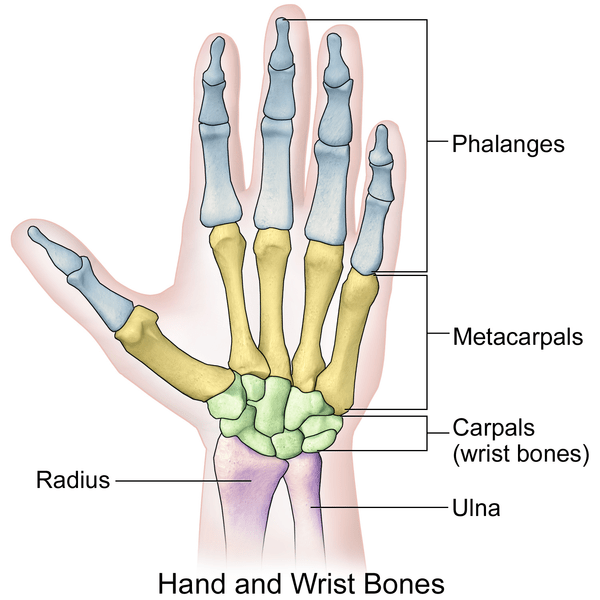
The MP joint is where the hand bone called the metacarpal meets the finger bones called the phalanges. The human hand consists of a broad palm metacarpus with 5 digits attached to the forearm by a joint called the wrist carpus. The 14 bones that are found in the fingers of.
A hand is a prehensile multi-fingered appendage located at the end of the forearm or forelimb of primates such as humans chimpanzees monkeys and lemursA few other vertebrates such as the koala which has two opposable thumbs on each hand and fingerprints extremely similar to human fingerprints are often described as having hands instead of paws on their front limbs. The front portion of the bone is thick and jagged to allow for its joining with other bones of the face. MP joints are important for both power grip and pinch activities and are where the fingers move with respect to the hand.
They are responsible for the fine motor functions of the hand. The eight bones of the wrist are. The intrinsic muscles of the hand are located within the hand itself.
About one fourth of the population does not have this tendon. For example so long to pinky here comes the thumb will help you with the carpal bone anatomy. The ulnar nerve enables us to grasp objects.
The scaphoid bone crosses both rows as it is the largest carpal bone. Not every hand surgeon uses the same words to describe hand anatomy and upper extremity hand and arm problems. The hand is composed of many different bones muscles and ligaments that allow for a large amount of movement and dexterity.
Anatomy of the Hand. These include the adductor pollicis palmaris brevis interossei lumbricals thenar and hypothenar muscles. The zygomatic bone is somewhat rectangular with portions that extend out near the eye sockets and downward near the jaw.
Each hand contains 27 distinct bones that give the hand an incredible range and precision of motion. This bone is on the thumb side of the hand near the radius. This bone rests between the scaphoid and triquetrum in the proximal row near the.
It results from imbalance between intrinsic muscles and comparatively weak extrinsic musclesContracture of the interosseous lumbrical or hypothenar muscles causes the fingers to stiffen and the. The rest have varying sizes of this tendon. The muscles in the forearm and palm thenar muscles all work together to keep the wrist and hand moving stable and well-aligned.
Hand Muscles and Hand Tendons. In this article we shall be looking at the anatomy of the intrinsic muscles of the hand. Consisting of the clavicle collar bone and scapula shoulder blade the pectoral girdle forms the attachment point between the arm and the chest.
Many surgeons have their own particular and preferential ways of describing normal anatomy and disease processes. The hand positioned at the end of the upper limb is a combination of complex joints whose function is to manipulate grip and grasp all made possible by the opposing movement of the thumb. It attaches to the wrist bone the pisiform and as well as the 5th hand bone.
The occipital bone houses the back part of the brain and is one of seven bones that come together to form the skull. Rotation of the radius around the ulna results in the supination and pronation of the hand. In a fracture of the scaphoid the characteristic clinical feature is pain and tenderness in the anatomical snuffbox.
An interactive quiz covering anterior view of Hand and Wrist Bones through multiple-choice questions and featuring the iconic GBS illustrations. Master wrist and hand anatomy with this simple carpal bones mnemonic. The hand also must be coordinated to perform fine motor tasks with precision.
The occipital bone is the trapezoid-shaped bone at the lower-back of the cranium skull. The hand can be considered in four segments. A single hand bone is called a phalanx.
The scaphoid is at particular risk of avascular necrosis after fracture because of its so-called. The thumb and large toe do not possess a middle phalanx. The scaphoid and the lunate are the two bones which actually articulate with the radius and ulna to form the wrist joint.
The carpal bone names can be labeled on an xray using mnemonics and acronyms. The red lines show. It travels along the elbow between the bone and overlying skin at the cubital tunnel.
It is located next to five of the cranium bones. The clavicle which gets its name from the Latin word for key is a long bone that connects the scapula to the sternum breast bone of the chest. The forearms ulna and radius support the many muscles that manipulate the bones of the hand and wrist.
However there are some pretty consistent terms labels and concepts that you should know when discussing your problem. These include both clinical and basic science studies along with case reportsSpecial features include Review Articles including Current Concepts and The Hand Surgery Landscape Reviews of Books and Media. The proximal intermediate and distal phalanges articulate with one another through interphalangeal joints of hand and interphalangeal joints of the foot.
The image below shows the bones of the hand from the back side. The palmaris longus tendon is a tendon with very little function in the hand. This is the.
In addition to reading this article be sure to watch our Hand Anatomy Animated Tutorial Video. Some biologists believe that the development of the human hand lead indirectly to the development of. The hyoid bone is a U-shaped bone that is held in place by the strap muscles of the anterior triangle of the neckThe bone has a central body forming the center of the U with two smaller protruding structures on the superior surface lesser horns and two larger bony protrusions from the body greater horns.
Carpal bones are a type of short bone shape and include s. The Journal of Hand Surgery publishes original peer-reviewed articles related to the pathophysiology diagnosis and treatment of diseases and conditions of the upper extremity. The hand is a complex part of the anatomy and there are numerous possible causes for nerve damage including traumatic injury repetitive stress infection.
The distal phalanges are the bones at the tips of the fingers or toes. Digits that extend from the palm of the hand the fingers make it possible for humans to grip the smallest of objects. There are 3 major types of bones in the hand itself including.
This complex structure connects the entire hand to the radius and ulna facilitates the passage of tendons together with the above mentioned neurovascular structures from the forearm to the hand and permits us to exploit all its movements.
Human Being Anatomy Skeleton Hand Image Visual Dictionary Online

Hand Bones Anatomy Structure And Diagram

Skeletal Anatomy Of The Hand Hand Clinics
Hand Pain And Problems Johns Hopkins Medicine
Hand Anatomy And Function Bone And Spine

Anatomy Hand And Wrist Bid Needham
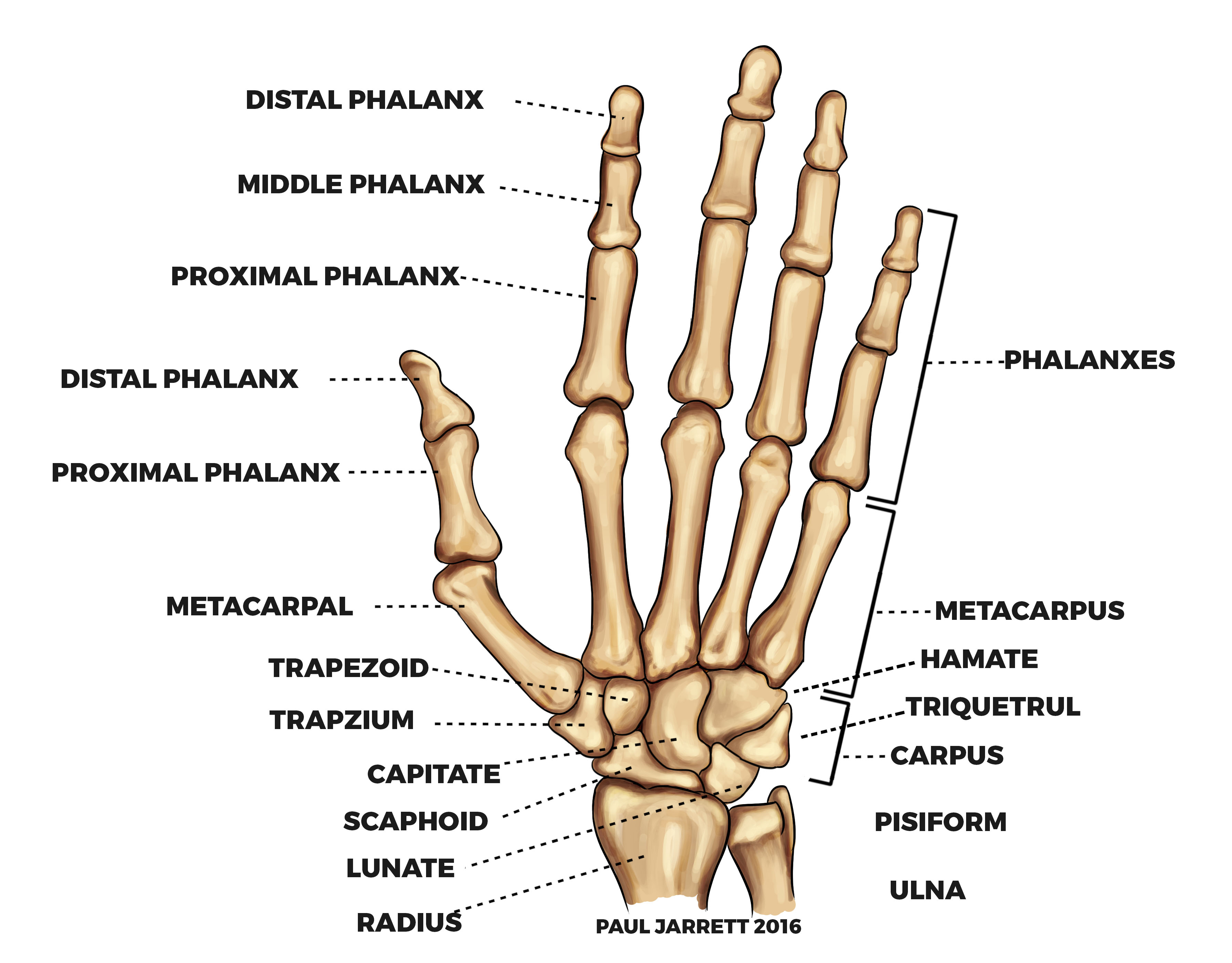
Mr Paul Jarrett Hand And Wrist Anatomy Murdoch Orthopaedic Clinic

Carpal Bones Wikipedia
0 Response to "Hand Bone Anatomy"
Post a Comment