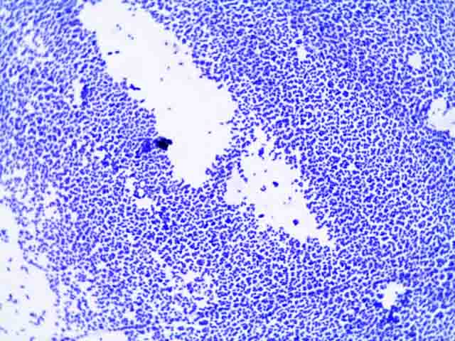
E Coli Simple Stain Results
Record this in the results section for this lab. Coli BL21DE3 cells and selected on carbenicillin LB agar plates at 30 o C.
Escherichia Coli Light Microscopy

Optical Microscopic Images Of E Coli Cells Following Crystal Violet Download Scientific Diagram

Simple Bacterial Stain Free Microbiology Images Photographs
EC-MUG Method for Determining E.

E coli simple stain results. Gram staining differentiates bacteria by the chemical and physical properties of their cell walls. The following method is a simple PresenceAbsence test that can examine 10-mL volume of juices. Bronchoscopy is a simple procedure that usually takes about 30 to 60 minutes.
Coli is a type of bacteria that can cause serious. Coli HCP impurities in products manufactured by recombinant expression. Data on the prevalence of ESBL fecal carriage remain scarce in Ethiopia.
Gram was actually using dyes on human cells and found that bacteria preferentially bind some dyes. Coli 6C01 the pKD46 plasmid with the λ red recombination genes and a temperature-sensitive replicon was transformed into chemically competent E. Methyl Red MR and Voges-Proskauer VP broth is used as a part of the IMViC tests as the medium in which both the Methyl Red and Voges-Prosakuer tests can be performed.
There are several excellent sites for designing PCR primers. And he describes how E. A isolation of a histidine-tagged green fluorescent protein GFP from a crude Escherichia coli lysate by affinity chromatography using an IMAC column.
The tube inoculated with Escherichia coli produces acid and gas. Colored pencils are available throughout the room. Background Extended-spectrum beta-lactamase ESBL producing bacteria present an ever-growing burden in the hospital and community settings.
A Best Book of the YearSeed Magazine Granta Magazine The Plain-DealerIn this fascinating and utterly engaging book Carl Zimmer traces E. WWW primer tool University of Massachusetts Medical School USA This site has a very powerful PCR primer. Writing should be simple and easy to understand.
Bacterial capsules are non-ionic so neither acidic nor basic stains will adhere to their surfaces. Purpose Procedure Results. Coli Escherichia coli is a bacterium that is typically found in a number of environments including various foods soil and animal intestines.
Coli in Shellfish Meats. Gram-positive bacteria and gram-negative bacteriaThe name comes from the Danish bacteriologist Hans Christian Gram who developed the technique. TEST PURPOSE REAGENTS OBSERVATIONS RESULTS Gram stain To determine the Gram reaction of the bacterium Crystal violet Iodine Alcohol.
Therefore the best way to visualize them is to stain the background using an acidic stain eg Nigrosine congo red and to stain the cell itself using a basic stain egcrystal violet safranin basic fuchsin and methylene blue. Last updated on May 22nd 2021. As a result the auxochrome enables the ionized chromogen to bind to cells or tissue fibres of opposite charge and thereby colour it.
Coli outbreaks have caused recalls or restaurant. Results of ColiComplete CC disc for E. Stain bacteria will either become purpleblue or pink during the procedure.
Sputum Gram Stain. Coli under the Microscope Types Techniques Gram Stain and Hanging Drop Method Introduction E. Coli is far more than just a microbial lab rat.
Gram-Positive Bacteria Explained in Simple Terms. Plasmid-containing cells were made electrocompetent and expression of recombination genes was induced with 01 arabinose. Colis hardiness versatility broad palate and ease of handling have made it the most intensively studied and best understood organism on the planetHowever research on Ecoli has primarily examined it as a model organism one that is abstracted from any natural history.
Medically reviewed by Alana. To generate marker-free atoB knockout E. Coli O157H7 are indole.
What do the results of a gram stain mean. Most patients are treated as outpatients but patients with pyelonephritis or prostatitis due to infection with E coli may benefit from hospitalization and supportive care. Rather it is a highly diverse organism with a complex multi.
Colis life and our own. Its genetic material consists mostly of one large circle of DNA 4-5 million base pairs mbp in length with small loops of DNA called plasmids usually ranging from 5000-10000 base pairs in length present in the cytoplasm. The tube inoculated with Enterococcus faecalis produces acid but no gas.
Colis pivotal role in the history of biology from the discovery of DNA to the latest advances in biotechnologyHe reveals the many surprising and alarming parallels between E. It is a simple broth that contains peptone buffers and dextrose or glucose. The bacterium was named after him since he isolated and found its components 2.
This kit was developed using broadly reactive polyclonal antibodies to the hundreds of different host cell proteins HCPs and is intended for use in determining the presence of E. Principle of Capsule Stain. Gram stain reagents Methyl.
Simple versus complicated infections location of infection susceptibility of strain adverse effects of antimicrobial agents and client compliance issues should be evaluated prior to treatment for E coli infections. Coli changes in real. It would be read as AG.
Coli colonies we first characterized the basic colony growth dynamics and morphology on solid M9 minimal medium which contained glucose and ammonium as the sole carbon and nitrogen sources respectively For all measurements performed with colonies including microscopy-based measurements the colonies. Therefore this study aimed to determine the prevalence of ESBL producing Escherichia coli and Klebsiella pneumoniae fecal carriage among children under five years in Addis. To investigate the spatiotemporal organization of metabolism inside E.
Metachromatic granules are also found in Yersinia pestis and Mycobacterium species. Theodor Escherich a German bacteriologist and pediatrician first discovered E. Spot growth from TSAYE plate to a filter wetted with Kovacs reagent.
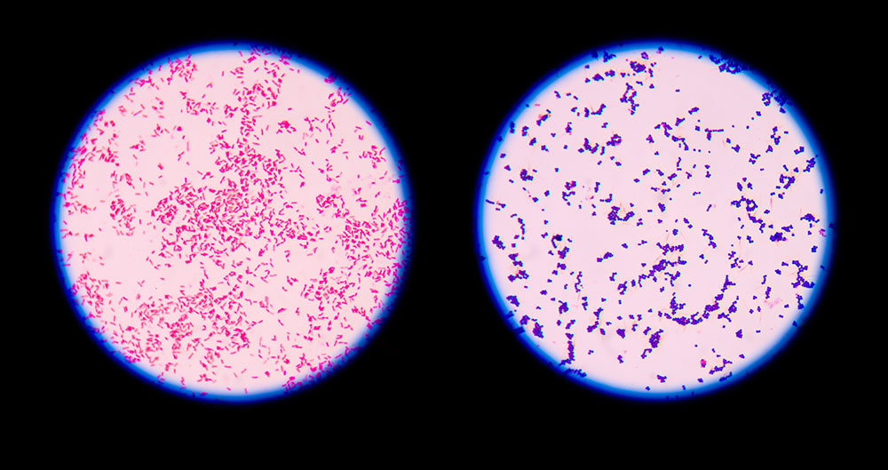
Escherichia coli is a tiny pink Gram- rod. The second part of the stain the auxochrome is a chemical group that ionizes the chromogen ie. Coli Commonly referred to as E.
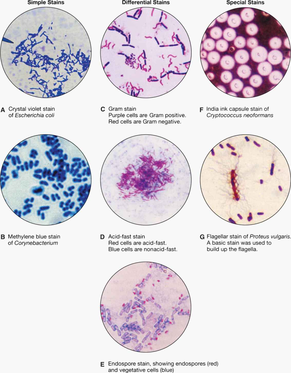
The Gram stain is a differential stain as opposed to the simple stain which uses 1 dye. All of the test results came out correct matching all the characteristic of E. After performing the Gram stain to determine that.
Albert stain is a type of differential stain used for staining high-molecular-weight polymers of polyphosphate known as metachromatic granules or volutin granules found in Corynebacterium diphtheriae. Stain-Free technology allows protein separation gel imaging and analysis in less than 30 min Visual confirmation of chromatography results using Stain-Free gels and imaging. It is named metachromatic because of its property of changing color ie.
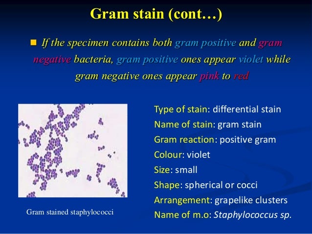
Staphylococcus epidermidis is a purple Gram sphere or coccus. Gram stain or Gram staining also called Grams method is a method of staining used to classify bacterial species into two large groups. It imparts a positive or negative charge to the chromogen group.
Our assay validation has. Draw a picture of a typical microscopic field and identify both Escherichia coli and Staphylococcus epidermidis. The bacterium you will be transforming Ecoli lives in the human gut and is a relatively simple and well understood organism.
As a result of the use of 2 dyes making this procedure a differential.
4 2 Specialized Bacterial Staining Techniques Biology Libretexts

Simple Staining Principle Procedure Results And Application Biology Ease

9 Gram Staining Best Practices Microbiologics Blog

Microscope Imaging Of Methylene Blue Stained E Coli Cells Harbouring Download Scientific Diagram

4 Principles Of Laboratory Diagnosis Of Infectious Diseases Basicmedical Key

Lab 3 Review Of Lab 2 Gram Staining Record Results On Pg 35 Gram Positive Purple Gram Negative Pink Bacillus Subtilis Escherichia Coli Klebsiella Ppt Download

Laboratory Perspective Of Gram Staining And Its Significance In Investigations Of Infectious Diseases Thairu Y Nasir Ia Usman Y Sub Saharan Afr J Med

Bacterial Staining
0 Response to "E Coli Simple Stain Results"
Post a Comment